Detalhes sobre a participação de 04 pesquisadores do CEPID BRAINN em um dos mais importantes eventos internacionais sobre epilepsia.
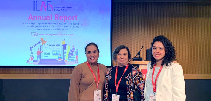
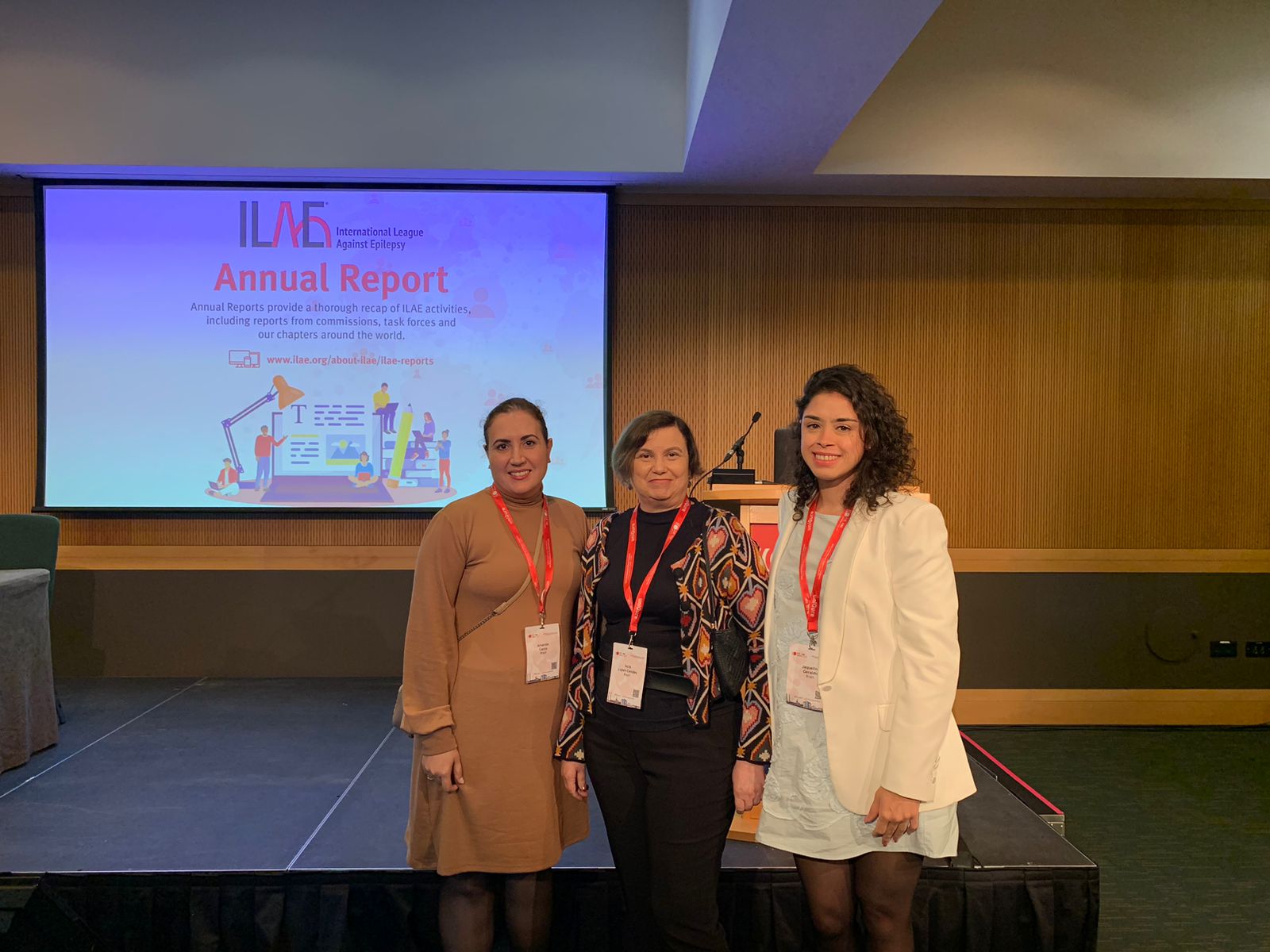
Os pesquisadores Fernando Cendes, Iscia Lopes-Cendes, Jaqueline Geraldis e Amanda Morato do Canto participaram do 35th International Epilepsy Congress (IEC), evento organizado pela International League Against Epilepsy (ILAE) e que este ano ocorreu entre os dias 02 e 06 de setembro na cidade de Dublin, na Irlanda. O evento contou com a participação de mais de 350 palestrantes de todo o mundo, distribuídos em mais de 120 sessões, abordando temáticas diversas sobre avanços científicos e médicos na epilepsia. Mais de 1300 trabalhos foram apresentados no total.
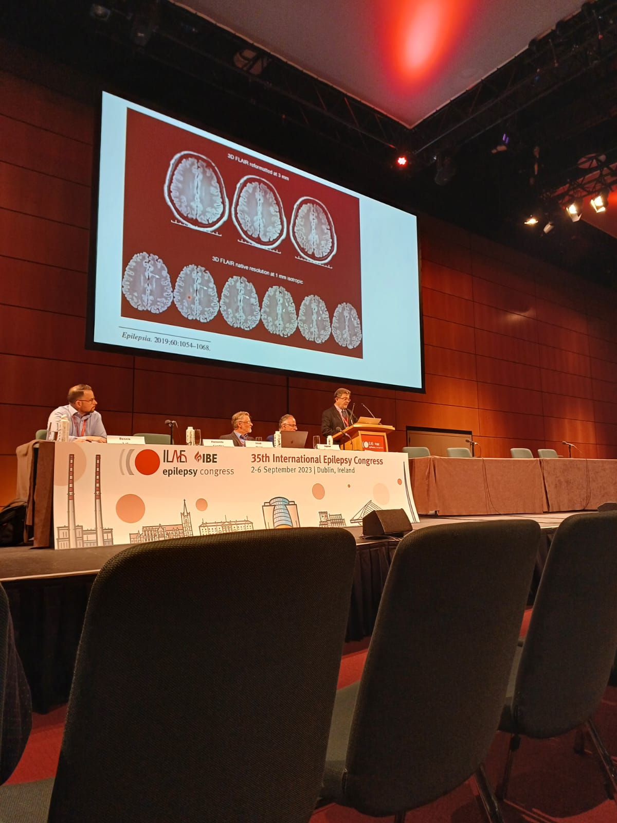
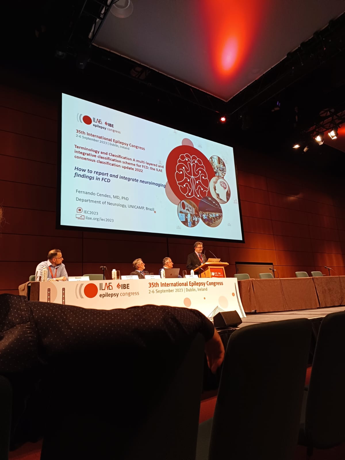
Fernando Cendes foi o speaker convidado para 02 sessões: uma palestra no sábado, dia 02, intitulada “Common epileptic pathologies: temporal epilepsy“, durante o curso “Neuroimaging in epilepsy – what the clinician should know“, e uma na quarta-feira, dia 06, com o tema “How to report and integrate neuroimaging findings of FCD“, durante o painel “A multi-layered and integrative classification scheme for FCD: the ILAE consensus classification update 2022“.
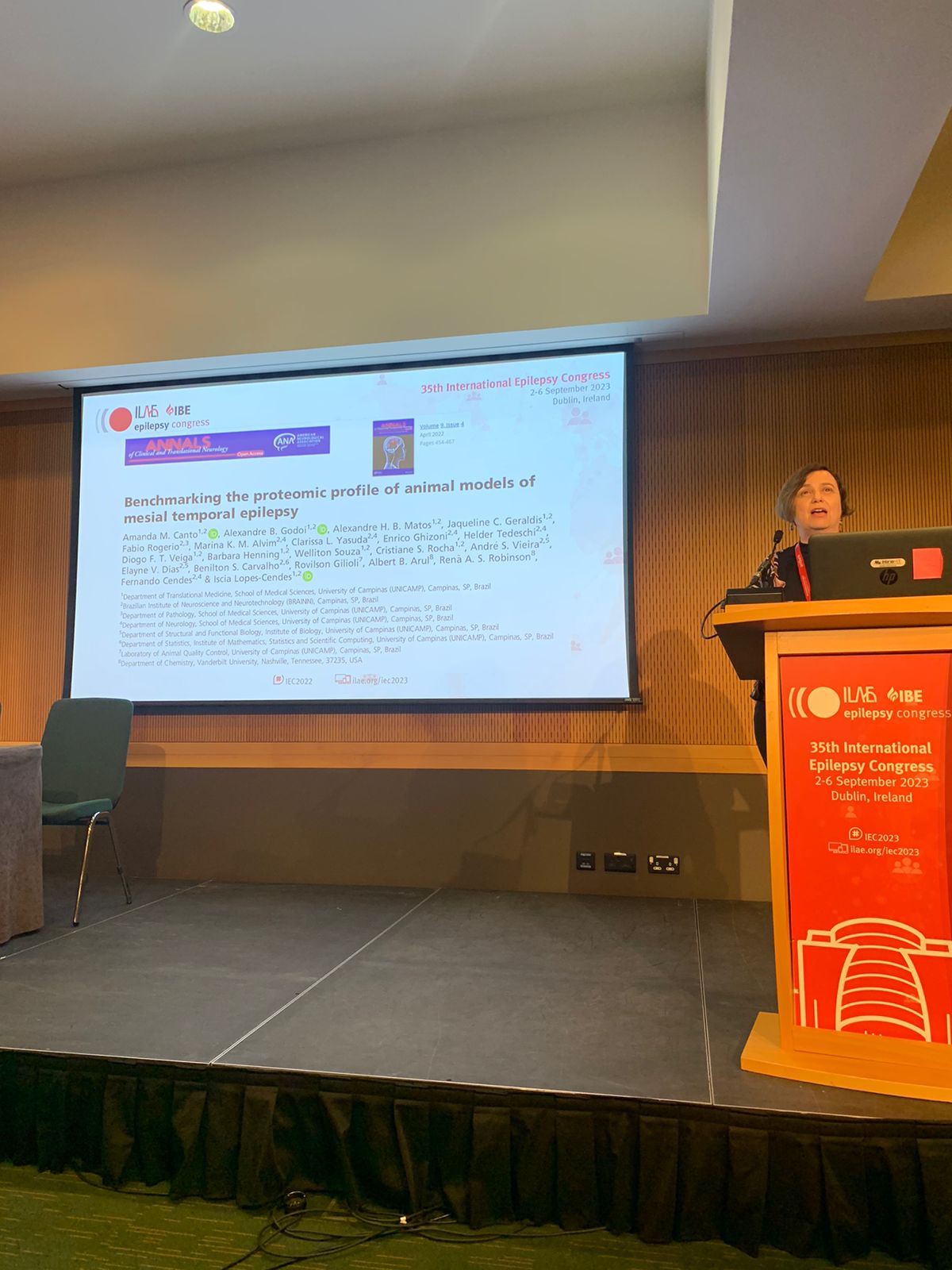
Iscia Lopes-Cendes apresentou o tema “Commonalities between multi-omics studies in basic and clinical epilepsy, do they translate?” durante o painel “Multi-omics in acquired epilepsies: beyond genes and transcripts“, que ocorreu no dia 05 de setembro.
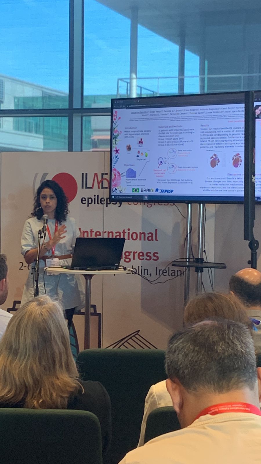
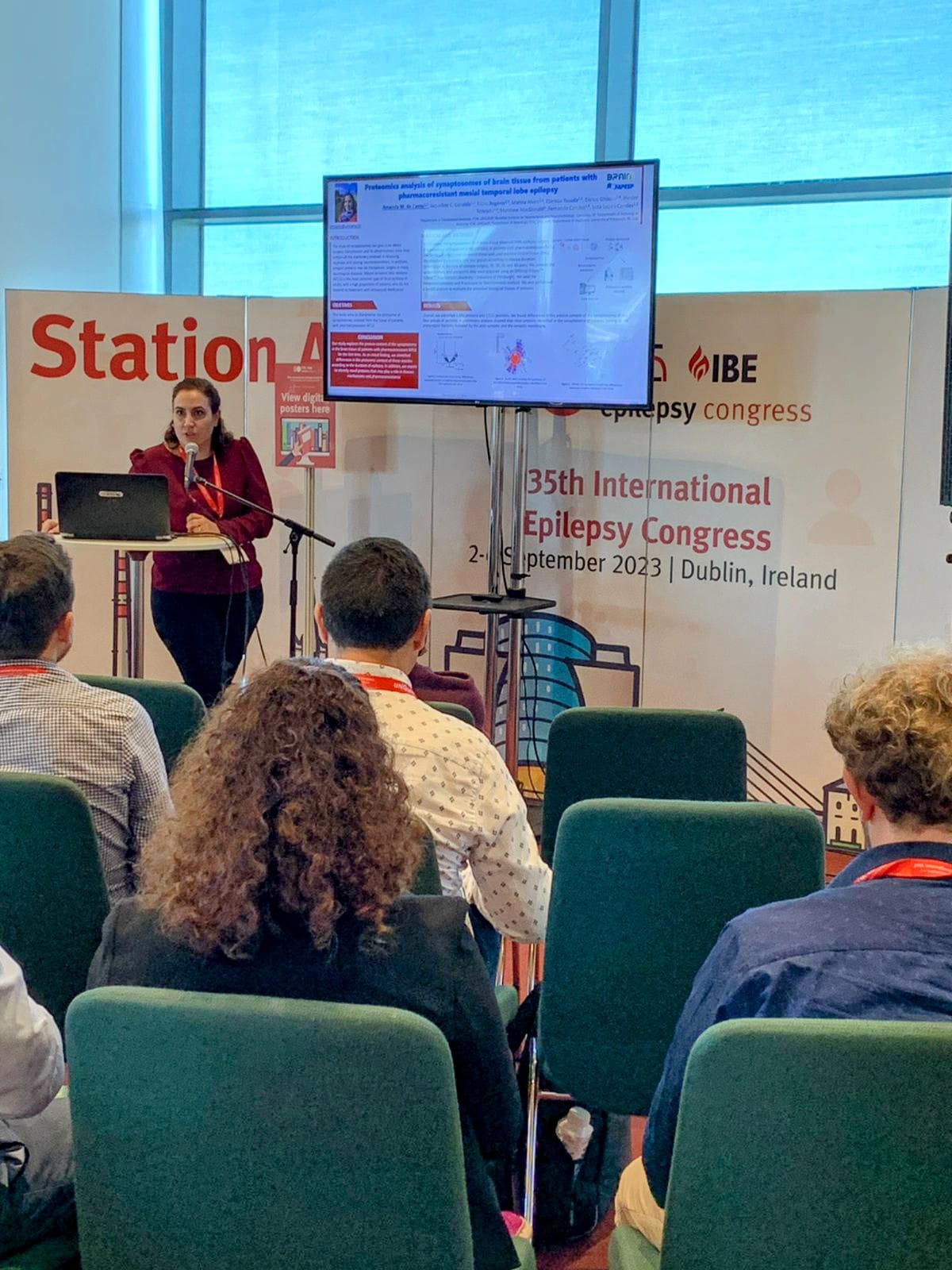
Amanda M. do Canto apresentou o trabalho “Proteomics analysis of synaptosomes of brain tissue from patients with pharmacoresistant mesial temporal lobe epilepsy” e Jaqueline Geraldis o estudo “Uncovering the multi-omic single-nuclei landscape of hippocampal sclerosis type I from patients with mesial temporal lobe epilepsy: clues into mechanism“. Veja detalhes sobre estes trabalhos ao final do post.
Confira a seguir fotos da participação dos pesquisadores do BRAINN no evento. Clique para abrir as imagens em alta resolução.
Detalhes sobre os trabalhos apresentados
Website oficial do evento
Relacionados/Related:
 Purple Day veste o mundo de roxo para conscientizar sobre a epilepsia
 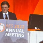 Fernando Cendes profere a ‘Lombroso Lecture’ durante encontro da American Epilepsy Society
  75th AAN Annual Meeting: trabalhos apresentados por pesquisadores do BRAINN
  Concurso fotográfico “Olhares sobre a Epilepsia” recebe inscrições
 English