

July 28th, 2015 Text by WebContent

MRI machine: revolutionizing clinical diagnosis since the 1970’s.
Magnetic Resonance Imaging (MRI) is a quite recent technique (the first prototype was completed in 1977), but has already revolutionized medical diagnostics. Through radio waves and magnetism – both painless and harmless to human beings – the technique is able to create detailed images of organs and tissues in three dimensions. It is widely used in the detection of various diseases such as cancer, stroke and multiple sclerosis. But for these images to be generated, it takes a lot more than just magnetism and radio waves …
Essential to medical exams such as MRI (and others that produce images of our body) is to organize the information acquired by the equipment and examine it in a computer to obtain an accurate and reliable diagnosis.
BRAINN researcher Roberto Lotufo, of the School of Electrical and Computer Engineering (FEEC) of Unicamp, and his research group have been working to facilitate and improve these image analysis processes. They are currently developing software that processes and analyzes brain images, also supporting clinical studies that uses MRI.
Next, some of Lotufo’s and his team main research lines.
STUDYING THE BRAIN IN 3D, WITH THE HELP OF BRAINN SOFTWARES
SKULL-STRIPPING

Brain imaging by MRI.
After a MRI exam, the first stage of a brain image processing is called Skull-stripping.
The technique of “skull-stripping” is used for this purpose. It is a computational method which enhances the results of MRI and allows the doctors to discern certain tissues and structures in higher resolution.
In this case, the software separates the skullcap from the brain tissues, allowing the analysis of all the other images. There are already Skull-stripping tools available, but Lotufo’s group is developing a new and better one – with it, the tests will be done faster and more accurately.
“Most current softwares have been validated with the study of less than 100 patients,” says Lotufo. “They usually have a high error rate and are very time consuming. For our software, we intend to use the studies of a thousand patients for validation. We plan to obtain results within a year”.
WHAT IS SKULL-STRIPPING?
A MRI exam obtains detailed data of various body tissues. The doctors, however, do not always want to study this whole set of information. When analyzing a patient’s brain, for instance, it is important to isolate the tissues which are of interest.
INTERACTING WITH BRAIN STRUCTURES
Lotufo’s group also works on the development of interactive software. The goal is to enable researcher to analyze MRI images in an innovative way.
“Our software allows the segmentation and the visualization of specific brain regions,” said Lotufo. “Each region can be individually selected and analyzed at different levels of black/white contrast”.
According to the researchers, the tool is more accurate and faster than others currently available. Moreover, it would also have the possibility of customization according to each study’s focus. More tests still need to be made, but the expectation is that the software will be available within the next six months.
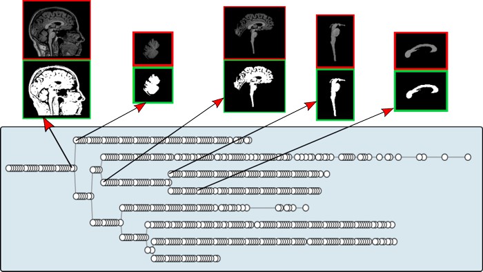
The interactive software developed by Lotufo’s group allows the user to isolate a specific brain region for analysis and then observe the region at different contrast levels. In the figure, each branch (or row) of the diagram represents a specific region and each circle a different level of contrast.
DETECTING INJURY
 Another line of research aims to develop software that can detect and characterize brain lesions. The methods currently available are for specific diseases only, so the group plans to create one that can be used for various conditions.
“This software would be ideal for patients who have multiple lesions in the brain which were caused by different diseases”, explains Leticia Rittner, professor at FEEC and BRAINN associated researcher. “For this software, we use a machine learning method. The machine is shown images of healthy brains and of brains that contain lesions, and is trained to identify and classify what is healthy tissue and what is not”.
MORE EFFICIENT CLINICAL STUDY = FASTER RESULTS
Lotufo and his group also seek to apply computational techniques to support clinical studies involving MRI.
The group collaborated, for example, with researches coordinated by Simone Appenzeller, from the Faculty of Medical Sciences of Unicamp. The study involved about 400 people and compared the brains of Lupus patients (see box below) with healthy brains to see if there were any differences in the volumes of brain structures. The researchers used a digital platform with several features that improved the use of medical data.
WHAT IS LUPUS?
Lupus is a chronic (or persistent) inflammatory disease. It affects about 250,000 people in Brazil, mostly women. It occurs when one person’s own immune system attacks tissues and organs of the body. It can affect different body structures such as skin, kidney, heart and lungs.
It is estimated that 50% of Lupus patients have their brain affected. In these cases, the disease can cause memory and cognition problems, confusion and fatigue. Although there is still no cure, there are already treatments that help to prevent and improve these symptoms. The sooner Lupus is diagnosed and treated, the better.
“We provide a platform, developed by us, that organizes data from clinical studies, processes and analyzes images and generates a final report”, says Rittner. “Thus, the researchers don’t have to worry about data analysis. With our platform, we facilitate the visualization of images, organize the information and deliver them the results”.
The platform is also being used for studies of Rheumatic Fever, coordinated by Appenzeller. The next step is to further disseminate the tool in academic studies.
WHAT IS RHEUMATIC FEVER?
Rheumatic Fever is a non-contagious, intense fever characterized by inflammation and acute joint pain. In most cases, it strikes young people. It is caused by inadequate treatment of infections caused by bacteria, particularly by streptococci.
“We are looking for researchers interested in this type of computer support to collaborate with new clinical studies in the brain imaging area,” says Lotufo.
The various research lines of BRAINN in the MRI area demonstrate how the correct configuration and analysis of data are fundamental to scientific and technological development. Through state-of-the-art software programmed in the CEPID’s labs, scientists and health professionals worldwide will be able to better understand the structure and function of the human brain – which is always the first step to create new treatments and cures for neurological diseases, providing greater quality of life for millions of people.

 Português do Brasil