
The study will obtain genetic information from children’s brain cells using innovative technologies and will be part of the Human Cell Atlas consortium’s Pediatric Networks.
BRAINN´s Success Stories – Innovative software allows more accurate diagnosis of the human brain
August 28th, 2015 Text by WebContent
A doctor receives the results of his epilepsy patient’s MRI exam. He opens the set of dozens of images on his computer and looks at them one by one, trying to find an injury or an abnormality. In one of the axial cuts, he notices a subtle, barely perceptible difference. Is that little darker spot responsible for the seizures? There is no way to be sure. Making a diagnosis like this one is difficult, even for specialized doctors. An error could pave way to a highly invasive, risky and unnecessary surgery.
With the intention of helping both patients that suffer from neurological disorders and health professionals, researcher Wu ShinTing – from the Faculty of Electrical and Computer Engineering (FEEC) at Unicamp and also a member of BRAINN – creates computer software that enables innovative views of MRI and PET (Positron Emission Tomography) exams. The computational tools developed by her group allow physicians access to a much larger amount of information, so they can make more accurate and informed decisions.
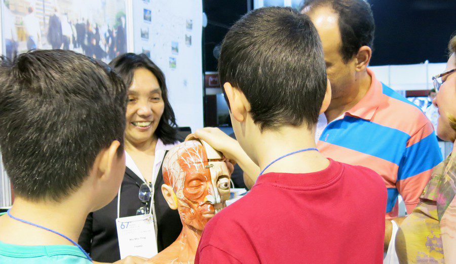
Researcher Wu ShinTing at SBPC Congress 2015 – research and education about the human brain.
INTERACTION TO OVERCOME CHALLENGES
Collaborations between Ting and the medical researchers began before the creation of BRAINN. About ten years ago, Ting presented her works to researcher Fernando Cendes, today BRAINN’s principal investigator. Cendes saw potential in looking at a three-dimensional images of the brain in a different way. Instead of the traditional straight layers, he would like to visualize them in curvilinear layers. Interested in the challenge, Ting began working on the development of a new algorithm.
After much research, months of studies and improvements, a first version of the program was completed in 2010. It was able to organize all the data and images from an MRI exam and provide the user with different views of the brain, straight or curvilinear, 2D and 3D.
“We realized that, depending on the angle, certain brain structures became more evident“, says Ting. “To facilitate medical diagnosis, our program offered multiplanar and curved cuts. Although there are other similar commercial tools available, our way of interaction is different”.
“Our bet is on interactivity – the researchers can move the cutting surface as they seem fit, either vertically/horizontally or diagonally, and observe brain structures from the angle they consider the best to confirm their assumptions”.
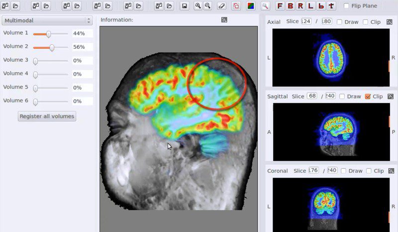
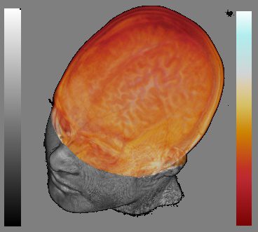
The computer program in action, combining magnetic resonance and PET-SCAN images. Click on the picture to watch a video about its operation.
AT BRAINN, INNOVATION NEVER STOPS
Four years later, a new version of the software – even more innovative – was developed by Ting’s group. The tool now included the visualization of PET exams aligned with MRI. Thus, the user could merge the anatomical images of the MRI with metabolic results obtained by PET.
More accurate diagnosis and improved brain scans have the potential to increase the quality of life of those living with neurological diseases.
“In addition to integrating PET with MRI, our second version offers more angles and a larger viewing area, providing a comparative analysis between the two cerebral hemispheres”, says Ting. “We also made it compatible with Windows, Linux and Mac”.
So far, the application has been primarily used in epilepsy patients. However, it has the potential to aid in the detection of brain tumors and other neurological diseases. Ting says that the next step is to integrate additional medical exams options, in order to provide doctors with more information and make their diagnosis as accurate as possible.
Our lines of research are mainly focused on the demands for computational tools that corroborate the doctor’s conjectures. We always pay attention to new proposals to investigate suspicious areas, so we would like to add exams such as SPECT (Single Photon Emission Computed Tomography), DWI (Diffusion MRI) and even EEG (electroencephalogram) to our tool”, says Ting. “Some of these projects have already started and are in progress, but we still do not have a prediction for when a new version of the program will be ready”.
If you want to know more about the history of the project, and view images and videos of the software created by Ting’s group, you can access its website by clicking the following link:
MTK ProjectBRAINN´s Success Stories – Neural Probes
July 15th, 2015 Text by WebContent
Neural probes are widely used today in neuroscience research and have many applications. These small needles, when inserted into the brain, allow the stimulation of specific regions and the recording of electrical activity. However, they are not yet produced in Brazil. With that in mind, one of BRAINN’s projects is to develop neural probes in the country for the first time.
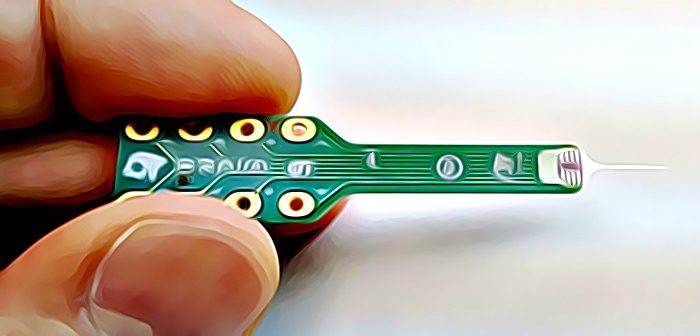
Neural probe created by BRAINN researchers, integrated into a circuit board that facilitates handling and allows for scientific uses.
“In 2010, researcher Hercules Neves invited me to be part of an European project of which he was coordinator, called Neuroprobes” says Roberto Panepucci, one of the researchers in BRAINN’s neural probes project. “The following year I came in contact with BRAINN, and we realized that developing neural probes here would be an interesting project.”
The activities began in 2012 with two master projects, supervised by Panepucci. Students André Malavazi and Jesus Arbey developed prototypes of the probes in the Renato Archer Information Technology Center (CTI), which have been tested with promising results.
PROBES MADE IN BRAZIL
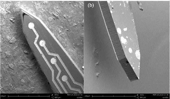
SEM images of the insertion shaft tip of a probe.
“During electrical activity recording tests stimulated by light pulses in live animals, our probes proved to be better than the ones already available on the market,” says Panepucci. “The silicon used in neural probes being sold today absorbs light and generates an electrical interference in the neural signal. Our probes are made from polymers, so this effect was significantly reduced, caused only by interaction with the metal”.
Besides their usefulness to scientific research, neural probes produced in the country could also bring economic advantages and might be easier to obtain.
“Currently, researchers working with neural probes in Brazil need to import them,” says Roberto Covolan, one of the project’s coordinators. “By producing these devices in the country, bureaucracies related to the importing process would be avoided. The cost of the probes would also be reduced”.
TECHNOLOGICAL INNOVATION AND CUSTOMIZATION
One of the challenges related to neural probes is data transfer. Since the probes have many electrodes, they need to be connected to many wires. This could make it difficult to carry out experiments and may limit the minimum size of the apparatus. “For these reasons, we are now working on the development of probes that can transmit information wirelessly,” says Covolan.
The next steps of the project involve making the neural probes available to several research groups that have shown interest in the equipment.
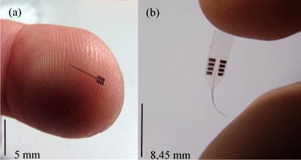
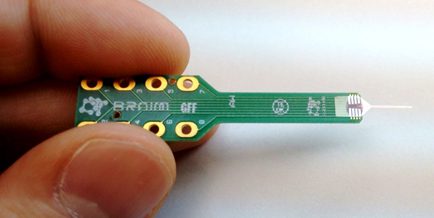
((left) Finalized neural probe produced by BRAINN. (right) Example of the equipment’s flexibility.
“One of the main advantages of developing probes here is the possibility of customization,” says Panepucci. “We created a software that lets you easily draw the probes and build a prototype from it. This way the probe can be made according to the demands of the researcher“.
André Vieira, postdoctoral student at FCM/Unicamp, is one of the scientists already interested in the neural probes. Vieira works with the molecular mechanisms of epilepsy, and part of his research involves the stimulation and the recording of brain activity in animals, particularly in the hippocampus area.
Vieira has conducted tests of mechanical nature with the equipment and noticed, for instance, that in order to use them it is necessary to remove the dura-mater, the outermost layer of meninges that surround the brain. According to Vieira, this procedure might bring advantages to the experiments, as the probe would be better fixed in the brain and would thus reduce the chances of tissue injury. And the advantages of the probes go on and on.
“With neural probes it is also possible to simultaneously record activities in different regions of the hippocampus, which is not possible with an electrode,” he says. “In addition, we can isolate the activity of individual neurons”.
The next step in Vieira’s project is to carry out electrical activity recording tests. For these he will use a custom-made neural probe, which will have a greater length I order to reach the hippocampus. “We intend to conduct these tests in the upcoming months,” he says.
BRAINN´s Success Stories – Light to See ‘Inside’ the Brain
November 9th, 2015 Text by WebContent
The patient has been hospitalized for a few days at Unicamp´s Hospital. He has suffered a stroke. After the initial treatments were done, the doctors are now closely following his progress. Has the stroke affected the man´s brain? Are the cognitive functions intact? To be sure, the medical team applies questionnaires and perform various tests – such as asking the man to move his arms and legs.
Even though they are efficient, all attempts to understand the consequences of a stroke are indirect. Doctors cannot directly check what is happening in the most important region of the body when it comes to stroke – i.e., the brain.
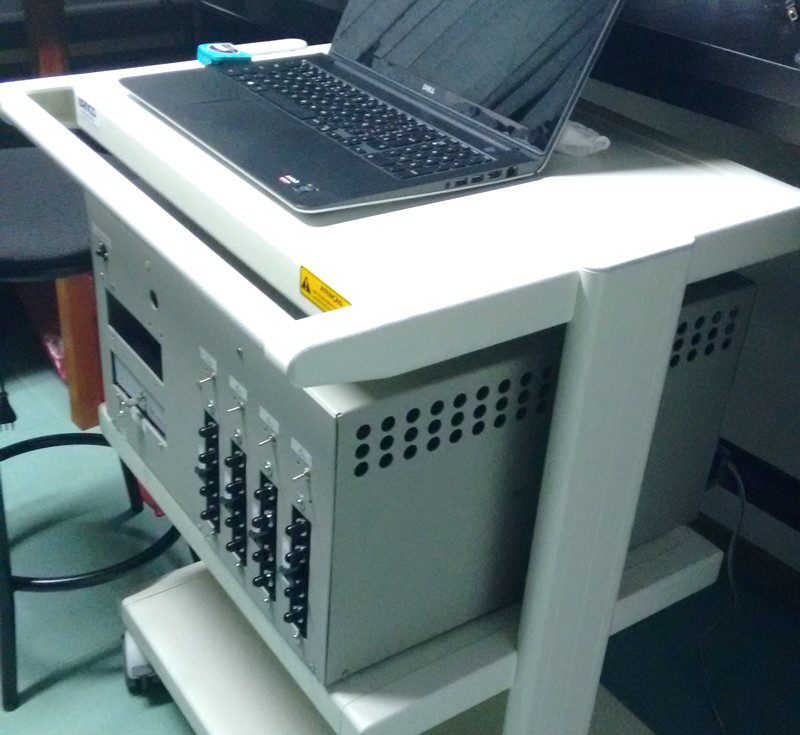
“To date, measuring the blood flow in stroke patients is an unsolved problem,” says Rickson Mesquita, researcher at the Neurophysics Group (GNF) at Unicamp and a BRAINN member. He and his research group are seeking an answer to this problem; an answer that is already being developed with the help of a modern device, unique in Brazil. Tests with this prototype may even reach the clinical phase later this year.
EQUIPMENT ALLOWS TO “SEE” WHAT HAPPENS INSIDE THE BRAIN
Light is an electromagnetic wave. The light that we can see, and which we call “colors”, is but a small part of a large electromagnetic spectrum that includes, for example, X-rays and radio waves.
The difference between what we can see and what we cannot see is the wavelength. We humans are able to see the part of the spectrum which is approximately between 400 and 700 nanometers (nm) long (a nanometer is one meter divided into a billion pieces).
The color red has a wavelength around 700nm. The near infrared is called so because it has a wavelength slightly higher, between 700 and 900nm – so we cannot see it.
To help diagnose patients who have suffered a stroke, Mesquita develops technologies based in near infrared light. In a simplified way, the light in the near infrared spectrum is able to pass through the skin and skull, interacting with hemoglobin molecules in our blood. This is important because hemoglobin is responsible for carrying oxygen to our entire body. Therefore, where there is hemoglobin, there is oxygen.
Knowing this, Mesquita´s idea was to develop a small device with lasers and infrared light detectors in order to use it in medical practice.
It works like this: the light is emitted by the device, enters our body, interacts with hemoglobin molecules and comes back to the detector. However, the intensity of light that returns is different from the original – the final intensity depends on the amount of hemoglobin with which it interacted. Looking at the difference between emitted light and detected light, it is possible to infer the presence of hemoglobin – and hence of oxygen – in the region under analysis.
“The device we developed is the first in Brazil that is able to measure blood flow in the brain microvasculature,” says Mesquita. “The greatest advantage of it in relation to the monitoring currently done in hospitals is that we can see, in real time, what is happening in the brain of patients who have suffered a stroke.”
“We can see, for example, if the brain of ischemic stroke patients are receiving blood and oxygen properly”, explains the researcher. “Moreover, it is possible to individualize treatment according to the specific characteristics of each patient we are seeing.”
The project, which began in 2013, had its first results at the end of 2014. The tests were done in healthy volunteers and confirmed the accuracy of the equipment. Now, the next step is to take the idea to hospitals and monitor stroke patients.
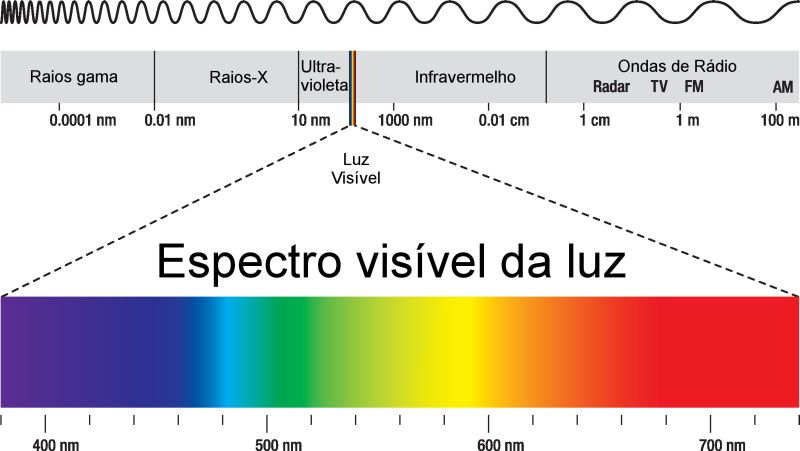
The light that we can see is only part of the electromagnetic spectrum. Note, in the above image, the infrared range.
TECHNOLOGY TO STUDY HEALTHY BRAINS
The device developed by Mesquita and his group can also be used to study healthy brains. One example is its application in research of brain networks, in which the goal is to understand how different regions of the brain are interconnected. The function of the device is to help determine which brain structures are active when certain activities and stimuli are present, since in these cases they have increased blood flow and consume more oxygen than regions not involved in the processes.
“The use of optical devices for brain networks studies has grown in the last five years,” said Mesquita. “They have advantages over magnetic resonance imaging, which can also be used for this purpose. Optical devices are more practical, portable and allow you to perform tests with patients standing and in more natural situations. ”
The ischemic stroke occurs when a region of the brain stops receiving blood. In this case, the blood vessel may be blocked, halting the flow of blood and oxygen.
The hemorrhagic stroke occurs when a blood vessel within the brain ruptures, causing blood extravasation and swelling in the region.
Stroke is the leading cause of death and disability in Brazil. Anyone who suffers a stroke, regardless of the type, needs immediate care to decrease the chances of sequelae. Some of the main symptoms of a stroke are: paralysis of one side of the body or face, difficulty in walking, impaired speech and language comprehension, visual impairment and mouth deviation to one side of the face. If you notice these symptoms, seek help immediately!

 Português do Brasil